Salmonella Microscope - Various Bacteria Cells In Microscope Streptococcus Pneumonia Pneumococcus Enterobacteriaceas Escherichia Coli Salmonella Klebsiella And Others Stock Photo Alamy : The second objective of this study was to investigate the biofilm formation of salmonella enterica serovar blockley (7175) in catfish mucus extract for 48 h at 22 °c on.
Salmonella Microscope - Various Bacteria Cells In Microscope Streptococcus Pneumonia Pneumococcus Enterobacteriaceas Escherichia Coli Salmonella Klebsiella And Others Stock Photo Alamy : The second objective of this study was to investigate the biofilm formation of salmonella enterica serovar blockley (7175) in catfish mucus extract for 48 h at 22 °c on.. For hyperspectral microscope, 3 µl of bacterial suspension from all. Blockley (7175) on a stainless steel surface were 4 log cfu/cm 2 and 5.5 log cfu/cm 2 in 15 μg/ml and 375 μg/ml of catfish mucus extract respectively after 48 h incubation at 22 °c. Causes food poisoning in man. Page 1 of 1 start over page 1 of 1. We have the compound microscope you are looking for!
We have the compound microscope you are looking for! Page 1 of 1 start over page 1 of 1. Taken by nova nanosem microscope magnification: Digital microscopes are great for large classroom computer combined instruction. Enterica is the type species and is further divided into six subspecies that include over 2,600 serotypes.
Figure 5 Evaluation The Surface Antigen Of The Salmonella Typhimurium Atcc 14028 Ghosts Prepared By Slrp from static-01.hindawi.com Thus, the extent of intercellular spread and ulceration of the epithelium is minimal. The objective of this study was to determine the growth and survival of salmonella enterica in the presence of high and low concentrations (375 μg/ml and 15 μg/ml) of catfish mucus extract at 10 °c and 22 °c for 63 days. We offer the best quality for the lowest price. When viewed under the microscope, salmonella bacteria, such as salmonella newport will appear as pink rods. The test employs several histidine dependent salmonella strains each carrying different mutations in various genes in the histidine operon. However, they take up the counter stain (safranin) and will appear reddish or pink when viewed under the microscope. After incubation for 72 h, cultures of A detailed look at the proteins of salmonella bacteria by researchers from the pacific northwest national.
Serotypes were spread on microscopic glass slides in the center approximately in the area of 20 x 20 mm followed by drying for 10 min in the
Salmonella under the microscope of proteomics. For instance, salmonella bacteria look alike under the microscope but can be separated into many serotypes based on two structures on their surface: E coli is a more common bacterium than salmonella which causes serious food poisoning in food. Coli) and the enterobacteriaceae spp. Taken by nova nanosem microscope magnification: When viewed under the microscope, salmonella bacteria, such as salmonella newport will appear as pink rods. Two types, salmonella enteritidis and salmonella typhimurium are the most common in the united states and account for half of all human infections. Pathogenic for rodents and other small animals. Page 1 of 1 start over page 1 of 1. Serotypes were spread on microscopic glass slides in the center approximately in the area of 20 x 20 mm followed by drying for 10 min in the Enterica is the type species and is further divided into six subspecies that include over 2,600 serotypes. More than 2,400 salmonella serotypes have been. Students can take images, videos, and more.
These types of bacteria prefer living in warm blooded animals. Environmental swabs can be tested for the presence of salmonella, and the results will help you determine your risk for contamination. By miranda waldron this image is of salmonella bacteria, a low kv was essential in order to visualise the surface detail of the bacteria, so the image was taken with the vcd (backscatter detector), using beam decelleration. Salmonella were initiated by adding 5 m l of the appropriate stock cultureto9mlofbpw(bd)inaglassdilutiontube(16by125mm) with a plastic cap. Systemic spread of the organisms can occur, giving.
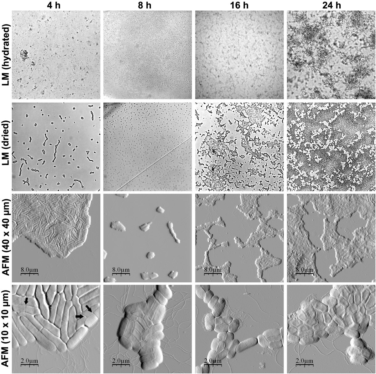
Roles Of Curli Cellulose And Bapa In Salmonella Biofilm Morphology Studied By Atomic Force Microscopy Bmc Microbiology Full Text from media.springernature.com Choose from 1,000s of microscopes. Salmonella (gram stain) gram negative rods 11. Pathogenic for rodents and other small animals. Thus, the extent of intercellular spread and ulceration of the epithelium is minimal. Bacteria are among the smallest, simplest and most ancient living. Morphology of salmonella typhi (s. The bar shown in the figure is equal to 1 μm. (a) these salmonella bacteria appear as tiny purple dots when viewed with a light microscope.(b) this scanning electron microscope micrograph shows salmonella bacteria (in red) invading human cells (yellow).even though subfigure (b) shows a different salmonella specimen than subfigure (a), you can still observe the comparative increase in magnification and detail.
The red shaped e coli is facultative anaerobic bacterium while its scientific notion is presented as escherichia coli or e.
Salmonella under the microscope of proteomics. Systemic spread of the organisms can occur, giving. E coli is a more common bacterium than salmonella which causes serious food poisoning in food. For instance, salmonella bacteria look alike under the microscope but can be separated into many serotypes based on two structures on their surface: Strains of salmonella cause illness such as typhoid fever, paratyphoid fever and food poisoning. Coli, do not escape the phagosome. Typhi under scanning electron microscope 10. Choose from 1,000s of microscopes. The bar shown in the figure is equal to 1 μm. Causes food poisoning in man. We have the compound microscope you are looking for! Statically grown bacterial suspensions were negatively stained and observed under electron microscope. Page 1 of 1 start over page 1 of 1.
When viewed under the microscope, salmonella bacteria, such as salmonella newport will appear as pink rods. Strains of salmonella cause illness such as typhoid fever, paratyphoid fever and food poisoning. After incubation for 72 h, cultures of (a) these salmonella bacteria appear as tiny purple dots when viewed with a light microscope.(b) this scanning electron microscope micrograph shows salmonella bacteria (in red) invading human cells (yellow).even though subfigure (b) shows a different salmonella specimen than subfigure (a), you can still observe the comparative increase in magnification and detail. It has all the major cell structures that can be found in prokaryotic cells, such as:
Confocal And Scanning Electron Microscopy Sem Analyses Of Salmonella Download Scientific Diagram from www.researchgate.net The bar shown in the figure is equal to 1 μm. More than 2,400 salmonella serotypes have been. Pathogenic for rodents and other small animals. Two types, salmonella enteritidis and salmonella typhimurium are the most common in the united states and account for half of all human infections. Students can take images, videos, and more. Typhi under scanning electron microscope 10. E coli is a more common bacterium than salmonella which causes serious food poisoning in food. Salmonella were initiated by adding 5 m l of the appropriate stock cultureto9mlofbpw(bd)inaglassdilutiontube(16by125mm) with a plastic cap.
It has all the major cell structures that can be found in prokaryotic cells, such as:
Systemic spread of the organisms can occur, giving. After incubation for 72 h, cultures of Till today, infections caused by various species and serovars of the genus salmonella. October 2nd, 2006 medgadget editors news. Statically grown bacterial suspensions were negatively stained and observed under electron microscope. Waldbusser january 6, 2010 biomapping may be the future to aid manufacturers in suppressing and controlling bacterial contamination The second objective of this study was to investigate the biofilm formation of salmonella enterica serovar blockley (7175) in catfish mucus extract for 48 h at 22 °c on. The bar shown in the figure is equal to 1 μm. Digital microscopes are great for large classroom computer combined instruction. Pathogenic for rodents and other small animals. E coli is a more common bacterium than salmonella which causes serious food poisoning in food. Choose from 1,000s of microscopes. We offer the best quality for the lowest price.
Blockley (7175) on a stainless steel surface were 4 log cfu/cm 2 and 55 log cfu/cm 2 in 15 μg/ml and 375 μg/ml of catfish mucus extract respectively after 48 h incubation at 22 °c salmonella. Get everyday low prices on our wide selection of premium microscopes.


0 Komentar